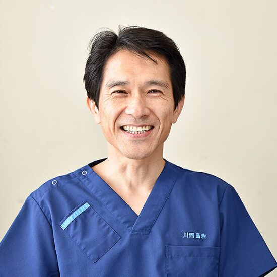
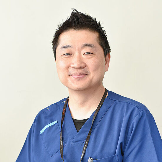
Masahiro Kawanishi ,M.D..Ph.D.
- Vice President, Chief of Neurosurgery
- Clinical professor of Osaka Medical College of Neurosurgery.
- Brain and spine surgery
Neurosurgery Services & Treatment
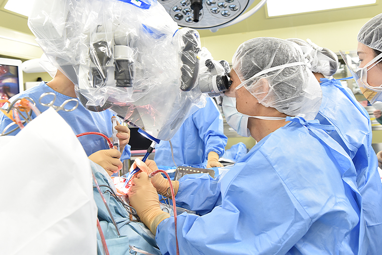
Our main goal is to help patients return home as soon as possible with as little burden as possible.
For the past 25 years, we have been providing appropriate treatment for each disease and each patient's situation by equating traditional craniotomy, spine and peripheral nerve surgery, and endovascular treatment with catheterization.
We are actively performing endovascular surgeries in which catheters are inserted into peripheral vessels in the brain and face, mainly through the femoral artery, without cutting the head as in conventional craniotomy, and have obtained excellent results even in very difficult cases. Using the latest angiography equipment, only one of which has been introduced in Kyoto, we are also able to provide treatment using some intracranial stents and liquid embolization materials, which are supplied only to facilities and surgeons. Diseases that can be treated by endovascular therapy are our first choice.
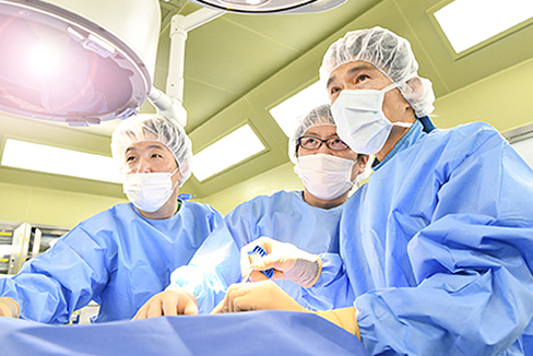
In the past, the treatment of the spine was to make a large incision, remove the affected area with a clear view, and fix it. However, our current treatment policy is to make the incision as small as possible and to use a microscope to repair the area with as little damage to the patient as possible.
Since the spine is a moving pillar of the body, our concept is to keep the area of removal as small as possible to avoid wobble. This reduces the need for screws to hold it in place. Even when it is necessary to insert screws, the treatment is performed with as little cutting of the muscles as possible. This is because back muscles are associated with back pain, and cutting them will prolong the recovery period. After spinal surgery at our clinic, patients are able to stand and walk from the next day and are discharged after stitches are removed. For compression fractures, which are common among the elderly, we perform “percutaneous vertebroplasty,” in which medical cement is injected into the bone, to relieve pain as quickly as possible and allow patients to return home as soon as possible, allowing them to leave the hospital the same day or the day after.
In this way, we aim to provide “minimally invasive” medical care that minimizes the burden on the patient and allows for a smooth return to society and home.
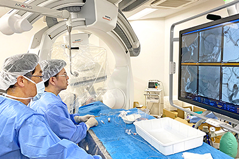
The main diseases that can be treated by endovascular surgery include the following.
Major diseases treated include brain tumors, spinal cord tumors, cerebrovascular disorders such as subarachnoid hemorrhage, unruptured cerebral aneurysm, cerebral arteriovenous malformation, intracerebral hemorrhage, cerebral infarction, head injury such as acute epidural and subdural hematoma, cerebral contusion, chronic subdural hematoma, spinal cord injury, cervical spondylolisthesis, cervical disc herniation, lumbar spinal canal stenosis, lumbar spondylolisthesis, compression fracture, lumbar disc herniation, and other spinal disorders, peripheral nerve diseases such as carpal tunnel syndrome, elbow tunnel syndrome, and functional disorders such as facial spasm and trigeminal neuralgia. Spinal disorders such as hernias, peripheral nerve disorders such as carpal tunnel syndrome and elbow tunnel syndrome, functional disorders such as facial spasm and trigeminal neuralgia, and other general neurosurgical conditions.
Percutaneous vertebroplasty (cement treatment) has been performed for spinal compression fractures since July 2003, and more than 1000 cases have been treated.It has excellent pain-relieving effects and can be performed as a one-day treatment.
https://www.e-neurospine.org/journal/view.php?number=1488
DOI: https://doi.org/10.14245/ns.2346936.468
What are the features of this machine?
In other words, in addition to “visualization,” simple and rapid “analysis” is possible, which reduces the burden on the physician performing the procedure and, by extension, has a positive effect on patient care.
In addition, the so-called “hybrid operating room” has been completed and put into operation with cleanliness control equivalent to that of an operating room. In the past 10 years, we have witnessed the advent of the “intracranial stent era,” and the structures are becoming more delicate.
The treatment of cerebral aneurysms with stents is becoming increasingly popular, and there are two main types of stents.
The general stent has a rough mesh structure and its main purpose is to act as a bulwark to prevent the coils to be embolized in the aneurysm from protruding into the mother vessel.
On the other hand, a flow diverting stent is a tool that reduces blood flow into the aneurysm and causes thrombosis by placing a stent with finer stents into the mother vessel.
Of course, not all aneurysms can be treated with all of these tools, as they are not suited to all patients.
When treating aneurysms with the FLOW DIVERTER, we are able to perform the procedure with the help of “Verify Now,” a testing device that can check platelet aggregation (blood thinning), and with an imaging device with operability and analysis capabilities, such as our department's angiography and fluoroscopy equipment (ARTIS icono D-Spin, Siemens). We are confident in our ability to provide “world standard” treatment to patients in the region.What are the features of this machine?
In other words, in addition to “visualization,” simple and rapid “analysis” is possible, which reduces the burden on the physician performing the procedure and, by extension, has a positive effect on patient care.
In addition, the so-called “hybrid operating room” has been completed and put into operation with cleanliness control equivalent to that of an operating room. In the past 10 years, we have witnessed the advent of the “intracranial stent era,” and the structures are becoming more delicate.

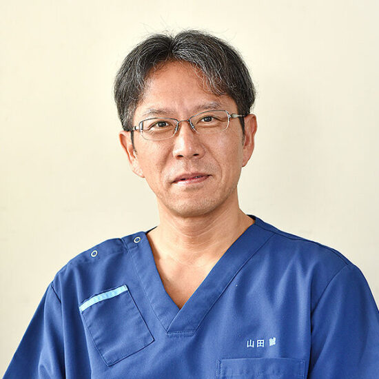
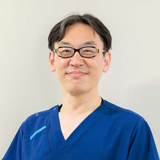
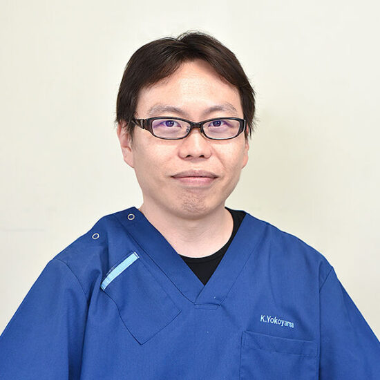
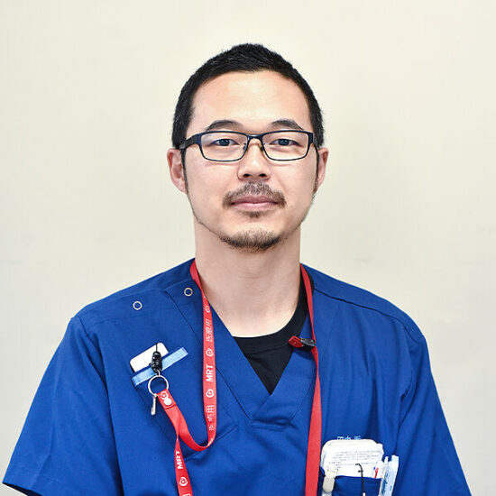

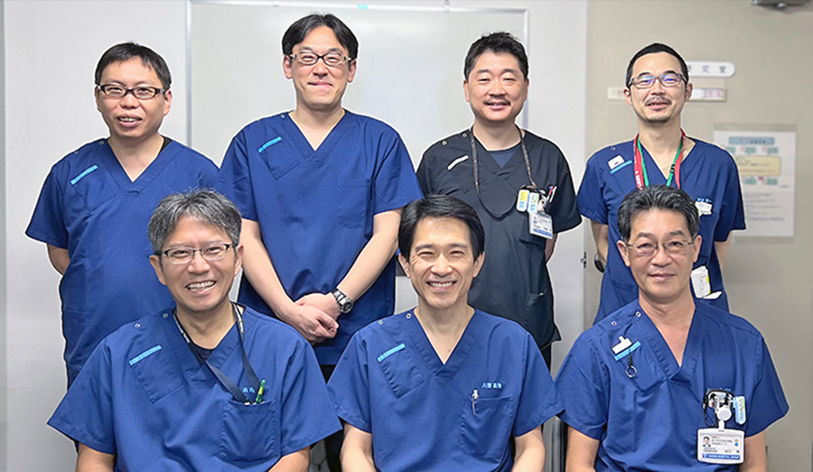
In order to improve surgical outcomes and techniques, the entire surgical process is recorded on video and digitized so that it can be published and evaluated by others through papers, academic conferences, and case review meetings with other institutions. We also use this information to provide guidance to junior surgeons and para-medical staff. All surgical cases are stored in a database so that they can be searched, tabulated, and published.
| working day | Mon-Fri. 9;00-12;00 |
|---|---|
| Address | 28-1 Ishidamori minamimachi Fushimiku Kyoto city Kyoto |
| Toll Free | 0120-72-6530 |
| Tel | +81-75-572-6530(Direct line) |
| Fax | +81-75-572-6276(Direct line) |
| Secretary | Tsujino SW |
| Access | Kyoto Metro Tozai line Ishida Sta. Exit 1. 5 minutes walk |
In order to improve surgical outcomes and techniques, the entire surgical process is recorded on video and digitized so that it can be published and evaluated by others through papers, academic conferences, and case review meetings with other institutions. We also use this information to provide guidance to junior surgeons and para-medical staff. All surgical cases are stored in a database so that they can be searched, tabulated, and published.
We receive criticism from others through publications and academic conference activities, and we conduct surgical training through autopsies of cadavers at other universities and in foreign countries for cutting-edge therapeutic acts, and we conduct animal experiments at our research institute. In addition, we train neurosurgeons who can handle craniotomy, spine/peripheral nerve surgery, and endovascular surgery as the three pillars of our education policy for future generations. We offer off-job training in microsurgery outside of the operating room.
Conferences, etc.
Our department participates in the following activities. For more information, please click below.
Stroke is classified into three types: cerebral infarction, in which a blood vessel in the brain becomes blocked; cerebral hemorrhage, in which a blood vessel breaks due to high blood pressure or other causes; and subarachnoid hemorrhage, in which a cerebral aneurysm ruptures. Advances in preventive medicine, mainly in lowering blood pressure and lipid management, have reduced the incidence of cerebral hemorrhage. However, subarachnoid hemorrhage has not decreased, and cerebral infarction is on the increase.
Stroke is the third leading cause of death after cancer and heart disease, killing about 130,000 people annually. It is the leading cause of death, accounting for nearly 30% of those who become bedridden. More than 250,000 new cases are diagnosed each year, due in part to the aging of the population and the increase in lifestyle-related diseases. It is estimated that the number of patients nationwide will reach 2.5 million by 2020, with approximately 60% requiring long-term care.
It is desirable to respond to stroke in a way that is appropriate for such a social background. In other words, an integrated and systematic response to stroke, from preventive treatment to acute, subacute, and chronic treatment, as well as home care, is considered necessary. To this end, it is essential to establish a team of medical professionals including physicians, nurses, rehabilitation therapists, and medical social workers, as well as cooperation with nearby rehabilitation hospitals, primary care physicians, and nursing care facilities.
It is desirable to respond to stroke in a way that is appropriate to this social context. In other words, an integrated and systematic response to stroke is considered necessary, from preventive treatment to acute, subacute, and chronic treatment, as well as home care. To this end, it is essential to establish a team of medical professionals including physicians, nurses, rehabilitation therapists, and medical social workers, as well as cooperation with neighboring rehabilitation hospitals, primary care physicians, and nursing care facilities.
A stroke care unit (SCU) is a specialized treatment ward for acute stroke patients where specialized medical staff provides intensive acute treatment and rehabilitation in an organized and systematic manner. The Stroke Treatment Guidelines (2009) verified that treatment in the SCU can reduce mortality, shorten hospital stay, increase the rate of discharge home, and improve long-term activities of daily living (ADL) and quality of life (QOL) of stroke patients (Grade A).
In order to respond to such social and community demands, the Department opened a 6-bed SCU that meets the facility standards specified by the Minister of Health, Labor and Welfare, and established the Stroke Center to better accept and treat acute stroke patients than ever before. Experienced neurosurgeons and neurologists, including stroke specialists, are available 24 hours a day. Patients with symptoms that indicate a high probability of stroke should contact us as soon as possible. Patients who have difficulty in determining whether or not they are suffering from a stroke should not hesitate to contact us.
The number of beds in the SCU and Neurosurgery Department is limited. Depending on the course of treatment and medical care, patients who require further rehabilitation may be transferred to a convalescent hospital through a regional cooperation path, or managed for transfer to other long-term care facilities or convalescent hospitals.
Our medical treatment is widely publicized through papers, video presentations, and academic conferences, both in English and Japanese.
ご連絡・アクセス方法